Hot key words: ENT Diagnostic series Otoscope Ophthalmoscope Penlight Hammer Tourniquet
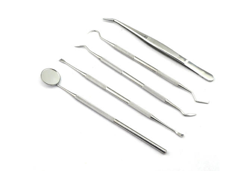
Let's learn about dental mirrors
- Categories:News Center
- Author:
- Origin:
- Time of issue:2021-02-02 11:10
- Views:
(Summary description)The emergence of oral endoscopes has brought a new model for oral examination and treatment. When the patient’s lesions are displayed in front of the patient, no more description or professional knowledge is required. Patients can also understand the urgency of treatment. Normal value: The oral cavity of a normal person is flat, smooth, smooth, and the oral mucosa is pink. Clinical significance: With the aid of clear and intuitive images, doctors can further discover the patient’s oral lesions and take various treatment measures in time. Abnormal results: abnormal signs caused by the disease, such as oral mucosa swelling, blisters, ulcers or spots Wait. People who need to be checked: abnormal oral color, pain, ulceration, smell, etc. Note: No special instructions. Note before the examination: Do not eat spicy, strong-tasting food. Meet the doctor's request. Inspection process: Use the storage function of the software to archive the patient’s oral situation to the greatest extent, which is conducive to long-term patient oral consultation. If supplemented by telephone return visits, it is more possible to retain long-term patients. The same as the medical clinic launched by 3D The speculum is equipped with a one-key capture function, which can instantly take color pictures and display them clearly. Related diseases: oral candidiasis, oral lichen planus, oral leukoplakia, tetracycline teeth, peritonsilar abscess, marginal gingivitis, chronic tonsillitis, tongue disease, leukoplakia, oral ulcers Related symptoms: Cannot quench thirst with drinking, sticky salivation, pus gums, oral erosions, gum chancre, white and moist fur, sputum bags in the mouth, sticky mouth, calculi, swollen gums
Let's learn about dental mirrors
(Summary description)The emergence of oral endoscopes has brought a new model for oral examination and treatment. When the patient’s lesions are displayed in front of the patient, no more description or professional knowledge is required. Patients can also understand the urgency of treatment.
Normal value: The oral cavity of a normal person is flat, smooth, smooth, and the oral mucosa is pink.
Clinical significance: With the aid of clear and intuitive images, doctors can further discover the patient’s oral lesions and take various treatment measures in time. Abnormal results: abnormal signs caused by the disease, such as oral mucosa swelling, blisters, ulcers or spots Wait. People who need to be checked: abnormal oral color, pain, ulceration, smell, etc.
Note: No special instructions. Note before the examination: Do not eat spicy, strong-tasting food. Meet the doctor's request.
Inspection process: Use the storage function of the software to archive the patient’s oral situation to the greatest extent, which is conducive to long-term patient oral consultation. If supplemented by telephone return visits, it is more possible to retain long-term patients. The same as the medical clinic launched by 3D The speculum is equipped with a one-key capture function, which can instantly take color pictures and display them clearly.
Related diseases: oral candidiasis, oral lichen planus, oral leukoplakia, tetracycline teeth, peritonsilar abscess, marginal gingivitis, chronic tonsillitis, tongue disease, leukoplakia, oral ulcers
Related symptoms: Cannot quench thirst with drinking, sticky salivation, pus gums, oral erosions, gum chancre, white and moist fur, sputum bags in the mouth, sticky mouth, calculi, swollen gums
- Categories:News Center
- Author:
- Origin:
- Time of issue:2021-02-02 11:10
- Views:
Scan the QR code to read on your phone
Latest News
-

2021-05-19
Otorhinolaryngology examination
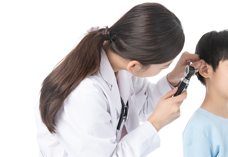
Otorhinolaryngology examination must be cautious because otorhinolaryngology is deep in the small cavity, so it is necessary to use special lighting devices and inspection instruments to check. There are 100 wattles with concentrated lens, such as check lights, frontal mirrors, otoscope, gas breathing otoscope, rifle forceps, cotton wool seeds, earwax hook, tongue depressor, anterior nasal mirror, posterior nasal mirror, indirect laryngoscope, tuning fork, sprayer and so on. read more
-
2021-02-02
List of medical equipment that needs to be purchased for ENT
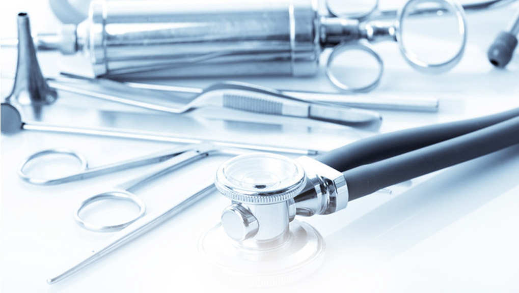
The Department of Otolaryngology includes the entire contents of ear, nose, throat, organs, esophagus, and related head and neck surgery. With the advancement and development of science and technology, various medical departments penetrate and promote each other, expanding the scientific scope of otolaryngology: To Ear microsurgery, ear neurosurgery, audiology and balance science, nasal endoscopy, nasal neurosurgery (nasal skull base surgery), head and neck surgery, laryngeal microsurgery, nasal neurosurgery, and the emergence of pediatric otolaryngology, greatly Enriched the scientific content of otolaryngology. To Configuration list of some ENT medical equipment: To Otoscope, medical magnifying glass, laryngoscope, rhinitis treatment device, tinnitus treatment device, periosteal treatment device, nasal irrigator, microwave treatment device, facial features checker, audiometer, medical headlight, carbon dioxide laser treatment device, hearing screening, Tonsillectomy instrument, ENT microscope, ion therapy instrument, pharyngitis rehabilitation therapy instrument, red light therapy instrument, etc. read more
-
2021-02-02
Related technologies of otolaryngology
Functional nasal endoscopy In the past, surgery for nasal polyps and sinusitis had to open up the nasal cavity and lift the face from the bottom up. The patient was severely injured, not to mention the pain, and the recovery was extremely slow; endoscopic microscopy was performed. After the operation, the minimally invasive effect greatly reduces the patient's pain, and also enables the patient to receive better, safer and effective treatment. The functional endoscopic nasal surgery carried out by the hospital is a brand-new technology. Its brightness is equivalent to 20 times that of a shadowless lamp, and a 3.5 mm hole can magnify the diseased tissue 500 times. Therefore, the patient’s diseased area can be seen by the doctor, and the clear vision enables the operation to achieve a more refined effect, and allows the operation to be carried out to the previously inaccessible area, thus transforming the previous traditional destructive surgery to the basis of completely removing the disease. , Functional minimally invasive surgery that preserves the physiological functions of the nasal cavity and nasal chamber as much as possible. Advantages of functional nasal endoscopy technology: ① Precise treatment: The brightness of the built-in cold light source is equivalent to 20 times that of a shadowless lamp. The clear vision enables the operation to achieve a finer effect. The 3.5mm hole can magnify the diseased tissue several times and transmit the synchronized image On the corresponding computer screen, the patient's diseased parts can be seen by the doctor, effectively saying goodbye to the past "blind man touching the elephant" era of blindly operating by hand and experience. ② Minimally invasive and less painful: The "American GLZ-40℃ minimally invasive rehabilitation technology" is usually used under nasal endoscopy, which is a high-tech treatment that uses low temperature (40~70℃) for tissue ablation. Compared with traditional laser microwave treatment with a temperature of up to 150°C, it greatly reduces tissue damage and patient pain. ③ Safe and fast: Automatically identify diseased tissues with high-intelligence probes to avoid excessive damage and reduce side effects. At the same time, an operation only takes a few minutes, safe and short, which is conducive to the recovery of patients after surgery. OPT optical fiber endoscope OPT fiber optic endoscope is composed of cold light source lens, fiber optic cable, image transmission system, screen display system, etc., with angles ranging from 0 to 120 degrees. Due to the use of laser illumination, the brightness is equivalent to that of traditional shadowless lamps. 30 times, and the lens diameter is only 2.7mm, it can easily pass through the narrow nasal cavity, nasal passage, and the internal structure of the ear canal to inspect the nasopharynx, ears, and even the internal structure of the sinuses, with matching surgical instruments It can also perform delicate treatment of ear, nose and throat diseases, so that the operation can reach areas that cannot be reached by traditional surgery, and realize the integration of non-invasive diagnosis and treatment of ENT diseases. Cryogenic plasma ablation As a minimally invasive surgical method for the treatment of snoring, low-temperature plasma ablation is mainly suitable for patients with nasal cavity and pharynx tissue stenosis and tongue hypertrophy, that is, obstructive sleep apnea syndrome. It maintains the safety of the local tissue structure and effectively reduces postoperative reactions (edema and pain). The treatment time is only 15-20 minutes, which can improve or effectively solve the problem of snoring at one time. Compared with traditional surgical methods, this technique mainly uses low temperature treatment, which directly acts on the submucosal soft tissues, does not damage the mucosa, has minimal damage, and has quick recovery and good results. Therefore, most people do not need hospitalization, as long as they are Just receive treatment in an outpatient clinic. The advantages of "low temperature plasma ablation": ① Short treatment time and no hospitalization: The treatment time is short, only about 15 minutes for one treatment, without hospitalization, and does not affect work and study. ② Precise positioning and low temperature ablation: Under the direct vision of the German STORZ nasal endoscope, magnify the diseased tissue 500 times for precise positioning and low temperature ablation. ③ Safe and no side effects: The unique disposable warhead can avoid cross-infection of iatrogenic diseases during the operation. It is pure and radiation-free and safer. ④ No surgery, no bleeding: no damage to the nasal mucosa, no scars, plasma therapy uses non-material contact, no nasal tissue damage, no surgery, no bleeding. ⑤ High curative effect, not easy to relapse: The recurrence rate of rhinitis patients is only two out of ten thousand by using American read more
-

2021-02-02
Let's learn about dental mirrors
The emergence of oral endoscopes has brought a new model for oral examination and treatment. When the patient’s lesions are displayed in front of the patient, no more description or professional knowledge is required. Patients can also understand the urgency of treatment. Normal value: The oral cavity of a normal person is flat, smooth, smooth, and the oral mucosa is pink. Clinical significance: With the aid of clear and intuitive images, doctors can further discover the patient’s oral lesions and take various treatment measures in time. Abnormal results: abnormal signs caused by the disease, such as oral mucosa swelling, blisters, ulcers or spots Wait. People who need to be checked: abnormal oral color, pain, ulceration, smell, etc. Note: No special instructions. Note before the examination: Do not eat spicy, strong-tasting food. Meet the doctor's request. Inspection process: Use the storage function of the software to archive the patient’s oral situation to the greatest extent, which is conducive to long-term patient oral consultation. If supplemented by telephone return visits, it is more possible to retain long-term patients. The same as the medical clinic launched by 3D The speculum is equipped with a one-key capture function, which can instantly take color pictures and display them clearly. Related diseases: oral candidiasis, oral lichen planus, oral leukoplakia, tetracycline teeth, peritonsilar abscess, marginal gingivitis, chronic tonsillitis, tongue disease, leukoplakia, oral ulcers Related symptoms: Cannot quench thirst with drinking, sticky salivation, pus gums, oral erosions, gum chancre, white and moist fur, sputum bags in the mouth, sticky mouth, calculi, swollen gums read more
-

2021-02-02
The correct use of the tongue depressor and 3 precautions
hongshun How to use the tongue depressor: 1. Open 50% GS 20 ml per tube every day and pour it into a sterile covered stainless steel tweezers tube for use. Replace the liquid and container twice a day. After use, the container is autoclaved and the tongue depressor is soaked in 500 mg/L chlorine-containing disinfectant for 30 minutes and then incinerated as medical waste. 2. When the child needs to use a tongue depressor to see a doctor, open the disposable tongue depressor package, soak the tongue depressor in 50% GS sterile solution for 1 minute, and take it out without dripping. Put it on the tip of the child’s tongue and let him taste the sweetness. Then let the child open his mouth wide and press down the root of the tongue quickly. At this time, the child will not cry or make trouble, and it will cooperate with the doctor’s various examinations. 3. In order to distract the children, you can hang bright murals and put some toys in the outpatient room to attract the children's attention and make the diagnosis quick. Using the tongue depressor correctly can reduce the fear of children and reduce doctor-patient disputes, whether it is for parents or medical staff, a good method can reduce a lot of trouble. Three precautions for the tongue depressor: 1. Pay attention to the disinfection of the tongue depressor; 2. Pay attention when pressing, don't let the child struggle uncomfortable; 3. The tongue depressor should usually be pressed on one third of the tongue. read more
-
2021-02-02
Otorhinolaryngology
General examination of the eye, including eye appendages and anterior segment examination. Ocular adnexa examination The ocular adnexa examination includes: examination of the eyelids, conjunctiva, lacrimal apparatus, eyeball position and orbit. Eyelid examination: usually inspection and palpation under natural light. Main observation: Whether there are congenital abnormalities of the eyelid, such as eyelid defect, blepharoplasty stenosis, ptosis, etc. read more
-
2021-02-02
Neurology
General examination of the eye, including eye appendages and anterior segment examination. Ocular adnexa examination The ocular adnexa examination includes: examination of the eyelids, conjunctiva, lacrimal apparatus, eyeball position and orbit. Eyelid examination: usually inspection and palpation under natural light. Main observation: Whether there are congenital abnormalities of the eyelid, such as eyelid defect, blepharoplasty stenosis, ptosis, etc. read more
Contact Us
Zhejiang Honsun Medical Technology Co., Ltd.
Add:Arts and Crafts Park, Beiao Industrial Zone, Sanjiang Street, Yongjia County, Wenzhou City, Zhejiang Province
Tel:86-577-88832888 86-18157711210
Fax:86-577-88870611
E-mail:jeashia@jeashia.com
Follow Us

Wechat QR code
Zhejiang Honsun Medical Technology Co., Ltd. 浙ICP备2021007239号-1 Powered by 300.cn


